The subaxial cervical, thoracic and lumbar vertebral bodies each have two ossification centres, which merge, and single ossification centres on each side of the vertebral arch (Fig. Chondrification of the CVJ begins at 45 days and chondrification of the C1 anterior arch begins at 50 to 55 days of gestation. The final steps in vertebral formation, chondrification and ossification occur after 6 and 9 weeks respectively. The ventral portion of the first cervical sclerotome forms most of the odontoid process. The axis is derived from both the fourth occipital and first and second cervical sclerotomes.

The posterior arch of the atlas is derived from both the first occipital and first cervical sclerotomes. Four occipital sclerotomes contribute to formation of the occipital bone, clivus and occipital condyles, anterior arch of the atlas and the apical, cruciate and alar ligaments. The craniovertebral junction (CVJ) is embryologically unique and complex. Underlying embryological processes include canalisation (secondary neurulation) and retrogressive differentiation. The sacrum and coccyx develop last at about 31 days of gestation from a large aggregate of undifferentiated cells (caudal cell mass) representing remnants of the primitive streak. After the notochord interacts with the overlying ectoderm to form the neuroectoderm, neural tube closure occurs to complete the process of primary neurulation. Since the notochord is created during gastrulation, multisystem abnormalities typically coexist with notochordal developmental anomalies. Sequence of appearance and fusion of the ossification centres as noted in the illustration d: Development of the C2 (axis): Six centres of ossification separated by four cartilaginous synchondroses. The sequence of appearance and fusion of the ossification centres as noted in the illustration. c: Development of C1 (atlas): The atlas develops from three centres of ossification separated by cartilaginous synchondroses-one for body and two for lateral masses. One of these loci eventually develops into the vertebral body while the other two develop into one half of the eventual vertebral arch.

One dorsal and one ventral primary ossification centre forms for each vertebral body these unite to create a centrum, which develops into three primary loci at the conclusion of embryonic development. b : Ossification of subaxial cervical, thoracic and lumbar vertebrae: This follows chondrification and typically occurs after 9 weeks of gestation. The six stages of vertebral development include: (1) gastrulation and formation of the somitic mesoderm and notochord, (2) condensation of the somitic mesoderm into somites, (3) formation of dermomyotomes and sclerotomes, (4) formation of membranous somites and re-segmentation with definitive vertebral formation, (5) vertebral chondrification and (6) vertebral ossification. The notochord expands and develops into the nucleus pulposus of intervertebral disks. Chondrification centres appear during the 6th week of development within a mesenchymal template, which enclose the notochord and developing neural tube. The sclerotomes migrate around the neural tube and the notochord forming the vertebral bodies, arches, transverse and spinous processes.

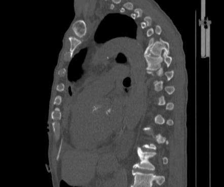
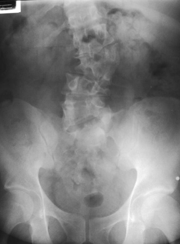
Accurate imaging interpretation of vertebral malformations requires knowledge of ageappropriate normal, variant and abnormal vertebral morphology and the clinical implications of each entity. These anomalies can occasionally mimic acute trauma (bipartite atlas versus Jefferson fracture, butterfly vertebra versus burst fracture), or predispose the affected individual to myelopathy. Malformations can be simple with little or no clinical consequence, or complex with serious structural and neurologic implications.

Malformed vertebrae can arise secondary to errors of vertebral formation, fusion and/or segmentation and developmental variation. A variety of structural developmental anomalies affect the vertebral column.


 0 kommentar(er)
